


.png)
.jpg)


Egyptian mummies come to life with Maltese AI scanner
University of Malta’s 3D visualisation scanner investigates animal mummies to study their materials in a non-destructive way

The University of Malta’s department of computer engineering is using AI to carry out 3-dimensional X-rays of Egyptian mummies.
ASEMI (automated segmentation of microtomography imaging) is a project led by Prof. Johann Briffa, head of the department of communications and computer engineering at the University of Malta.
Using artificial intelligence techniques, the tool provides microtomography images of Egyptian mummies. Microtomography works just like a medical scanner, using synchrotron radiation instead of a conventional X-ray source, to capture volumetric scans at much higher resolution and quality.
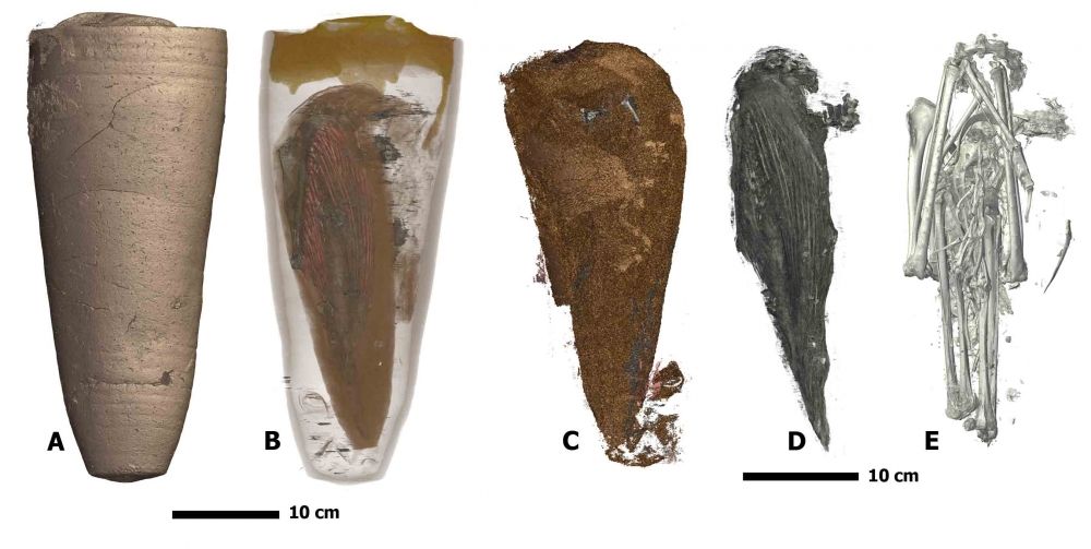
3D scan of an Ibis jar
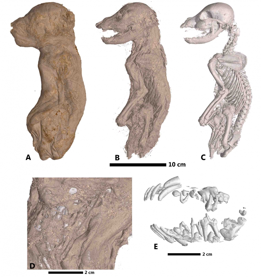
3D scan of a dog
This allows a 3D visualisation of the structure of materials in a non-invasive and non-destructive way, with applications in cultural heritage, materials research, life sciences, biomedical research and paleontology.
One of the most recent applications is in Egyptology, through the investigation of animal mummies. In this application, trained specialists manually segment the volume into the various component materials, such as textiles, organic tissues, balm resin, ceramics, and bones.
Depending on the complexity and size of the dataset, this process is very time consuming, typically taking several weeks for a small animal mummy.
With the construction of the new beamline BM18, the same process is expected to be applied to human mummies, which would take considerably longer.
The project was in partnership with the European Synchrotron Radiation Facility (ESRF) in Grenoble, France, which has developed unique expertise in the application of synchrotron imaging to paleontology and archaeology.
The main result of the ASEMI project is the development of a tool to automatically perform this laborious process. Using the developed software, the human specialist only needs to manually segment a small sample of the volumetric image. This is used to train and automatically optimise a machine learning system, which can then segment the whole volume in a fraction of the time previously required.
The accuracy obtained by the ASEMI segmenter approaches the results of off-the-shelf commercial software using deep learning, at a much lower complexity.
Following the principles of “Open Innovation, Open Science, Open to the World”, the developed algorithms, data sets, and results have been made freely available to the general public.
